The development stages of the embryo can be divided on the three trimesters that a pregnancy has, each of them is characterized by a group of phenomena:
–First semester: very fast cellular divisions, establishment of the germinal layers and the organogenesis begins
–Second semester: the organogenesis is fully completed
–Third semester: growth of the fetus and establishment of the functionalities of each organ.
Fertilization
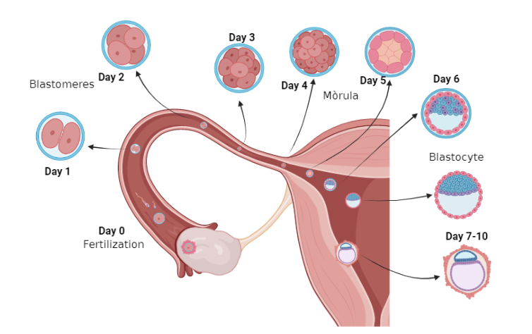
At fertilization, the egg contributes with 23 chromosomes, the organelles and all the machinery and food reserves while the sperm contributes with 23 chromosomes and the centriole. This fertilization process takes place in the Fallopian tubes that connect the ovary to the uterus, if it occurs later it will be a fertilization that will not lead to a pregnancy. This process is further explained in the “fertilization” post.
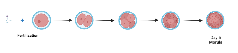
Once the egg is fertilized (Day 0) it is called a zygote which will activate its metabolism by activating segmentation. The embryo segments into different cells very quickly (1 division per day) without growth so the cells are smaller and smaller, that is to say, it goes from having one giant cell (the egg) to 32 “standard” size cells when it reaches the “morula” stage. This is thanks to the fact that all the necessary components are already in the cytoplasm of the egg and it is not necessary to generate more.
Fertilization occurs very close to the ovary, if it occurs later it will no longer trigger pregnancy.
The cells of the zygote are called blastomeres and they divide successively in a mitotic way while the embryo descends through the fallopian tube until it reaches the uterus on day 6. At this point, the zygote is called a morula since its shape is reminiscent of blackberries, it has 32 cells.
Implantation
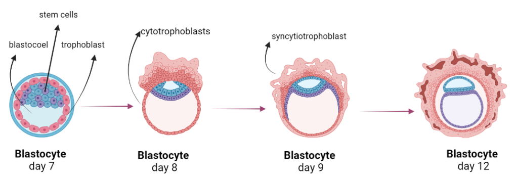
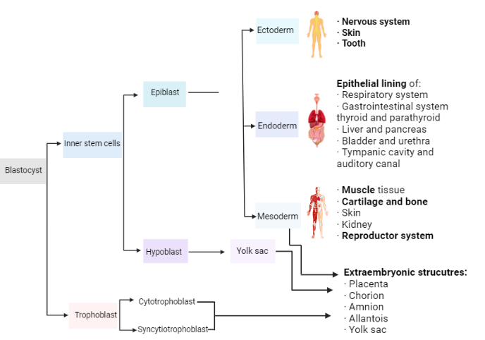
Up to this point, we have only seen division and movement of the zygote into the uterus. However, upon reaching the morula stage, cavitation occurs where the cells have already begun to differentiate. Part of the cells go to the periphery and will go on to form the trophoblast, which pumps a liquid called blastocoel inside, thus generating the blastocyst. There are some cells inside, they are pluripotent stem cells that will give rise to the new organism.
We found 3 types of stem cells in our body:
Tutipotent stem cells: capable of creating any sort of cells, both from our body and also placenta and amniotic sac.
Pluripotent stem cells: capable of creating any cell of the body.
Multipotent cells: are the ones more commonly found on our body and are the ones in charge of regenerating our tissues, this are capable to create a group of cells for example all the cells from the blood come from the same multipotent stem cell.
From this moment, on day 7, the zygote contacts the cells of the wall of the uterus (endometrium) and begins to implant itself inside the wall of the uterus. At the same time the cells continue to proliferate.
The contact causes the trophoblast to divide in two parts: the cytotrophoblasts and the syncytiotrophoblast. On the one hand, the cytotrophoblast are the internal cells from the trophoblast which are in charge of the nutrition of the inner stem cells. On the other hand, the cells from the exterior start to proliferate without cytokinesis (it becomes a mass of nuclei with a single cytoplasm, kind of like a bag full of nuclei) creating the syncytial trophoblast which penetrates the walls of the endometrium while the cells from the interior trophoblast feeds the cells of the internal mass. As they enter the wall of the endometrium, gaps and villi are created, these structures will participate in the formation of the placenta.
The syncytiotrophoblast is the placental barrier between maternal and fetal blood that allows exchanges in nutrients and gasses and also represents the endocrine tissue of the human placenta.
Gastrulation
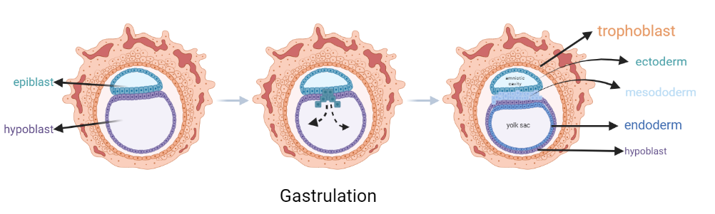
During this process, pre-gastrulation, we observe that the cells of the internal cell mass have differentiated into two layers of cells, the epiblast which will develop into both embryonic and extraembryonic tissue, and the hypoblast which will only form extraembryonic tissue.
Once the implantation is completed, gastrulation begins. This phenomenon is characterized by the migration of epiblast cells to the hypoblast generating a gap/path. The cells from the epiblast that cross the path will end up being endoderm and mesoderm while those that do not cross will generate the ectoderm.
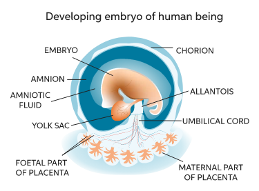
During this process of gastrulation the extraembryonic structures are starting to be created from the trophoblast, hypoblast and some cells from the mesoderm. This structures will participate in the protection of the new organisms, alimentation and interaction with the mother:
Chorion: layer that covers the embryo.
Amnion: protects the embryo creating the amniotic sac.
Allantois: in charge of the nutritional exchange with the mother, it connects the embryo with the umbilical cord.
Yolk sac: stores a reservoir of nutrients to nutrish the embryo in the first steps of development when the mother is still not connected to the child. After the connection is done it disappears since this function is performed by the placenta.
Organogenesis
After gastrulation, neurulation and organogenesis occur. The ectoderm closes the gap/path creating the neural tube from where all the nervous system is generated. From here all the organs of the body are as well created.
Besides, from the 25th day, the placenta begins to form, allowing the exchange of nutrients between the mother and the embryo. The embryo is no longer dependent on the nutrients from the egg. This is done through the umbilical cord which contains two arteries and a vein that carry blood to the placenta where gas and nutrient exchanges take place between the two bloods (the mother’s and the embryos’).
So, to make it clear here is a list of where does each part of our body comes from regarding this early embryo:
By week eight, organogenesis is complete. The fetus appears human-like and is prepared to undergo further growth and differentiation. So yes, the most complex part of the pregnancy often happens before mothers realize that they are pregnant.
But… if by eight weeks the organs are already created, what needs to be done until week 40?
Maturation
Basically what happens here is that each organ fully develops and correctly forms. Not only that but also systems are created: lungs are connected to the heart through the pulmonary artery, the small intestine connects with the colon, etc.
Some organs will also develop their function during this month and mothers will not fully develop until birth.